Specialized
Diagnostic Tests
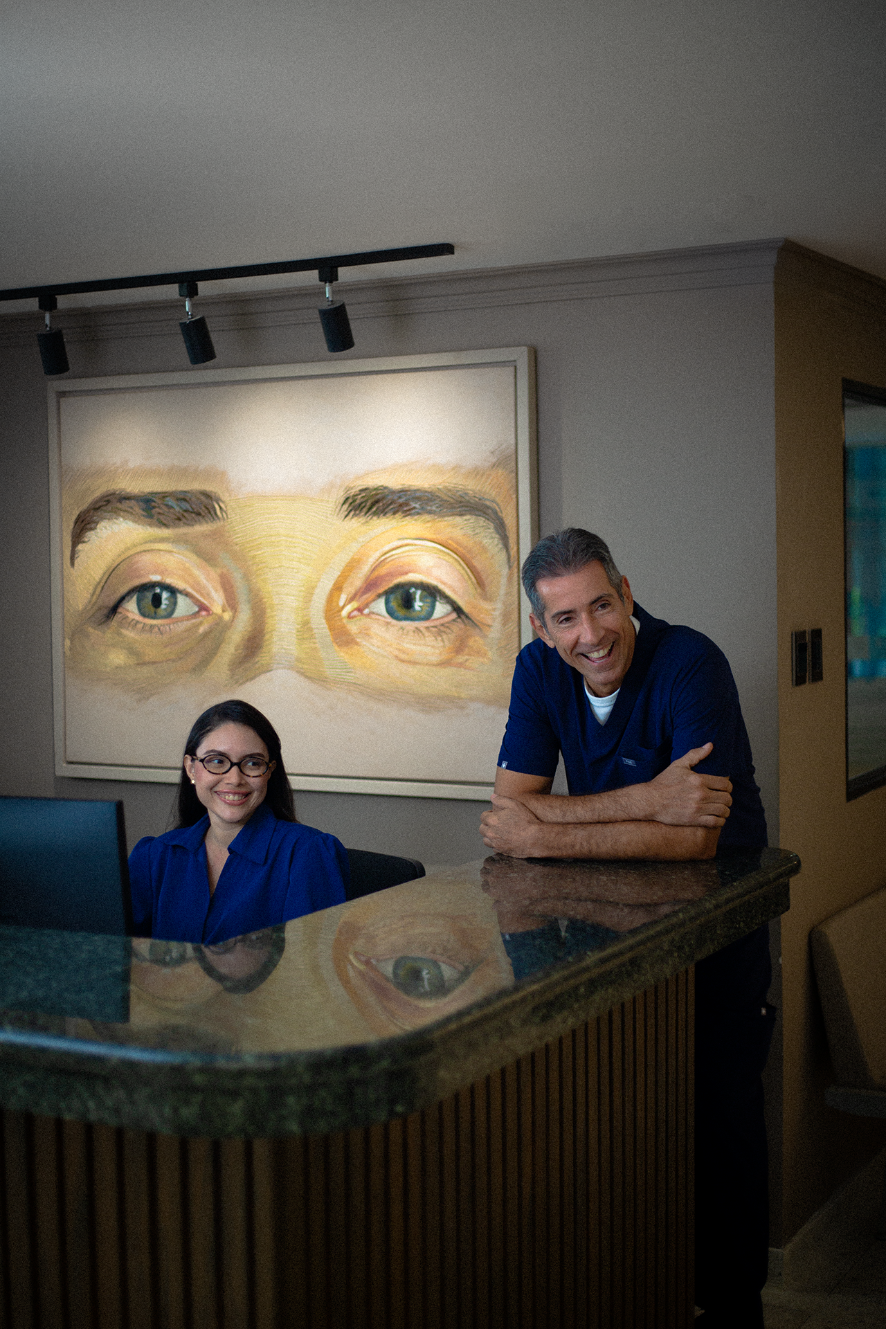
We put our soul into your eyes.
More than 25 years of experience.
High-Precision Visual Diagnosis with Advanced Technology
ANGIOGRAPHY - OCT
Imaging exam that combines optical coherent tomography with blood flow analysis, useful for detecting vascular alterations in the retina and choroid without the need for contrast.
RETINAL ANGIOGRAPHY
It uses intravenous dyes to evaluate retinal and choroidal circulation, detecting leaks, occlusions, neovascularization, or damage from diabetes and other vascular diseases.
OCULAR BIOMETRY
Measures with high precision the eye’s axial length, anterior chamber depth, and other dimensions—essential for planning cataract surgeries and intraocular lens implantation.
REFRACTIVE BIOMETRY
Calculates the eye’s dioptric power and predicts postoperative refraction. A fundamental tool for the personalized selection of intraocular lenses.
IOP CURVE (INTRAOCULAR PRESSURE CURVE)
Serial evaluation of intraocular pressure throughout the day to diagnose and monitor glaucoma, or to detect variations that may not be evident in a single measurement.
OCULAR ULTRASOUND
Ultrasound method that allows visualization of the internal structures of the eye in the presence of opacities. Useful for examining the retina, vitreous, or intraocular tumors.
UBM ULTRASOUND - ULTRABIOMICROSCOPY
High-frequency ultrasound that provides detailed images of the anterior segment of the eye. Essential for diagnosing glaucoma, tumors, and anterior chamber conditions.
ELECTRORETINOGRAM (ERG)
Measures the retina’s electrical response to light stimuli. Key for diagnosing hereditary, degenerative, or toxic retinal diseases.
ELECTROOCULOGRAM (EOG)
Evaluates the electrical potential between the cornea and retina during eye movements. It allows the study of the retinal pigment epithelium and light adaptation functions.
ELECTROPHYSIOLOGY EQUIPMENT
Set of devices used to perform functional tests such as ERG, EOG, and visual evoked potentials, which evaluate the bioelectrical functioning of the visual system.
VISUAL FIELD STUDY
Measures the sensitivity of central and peripheral vision. Essential for the diagnosis and monitoring of glaucoma, neurological lesions, and retinal diseases.
COLOR FUNDUS PHOTOGRAPHY
Detailed imaging of the retina, macula, or optic nerve to monitor and compare changes in conditions such as AMD, glaucoma, or diabetic retinopathy.
EXTERNAL OCULAR PHOTOGRAPHY – PANFLEX
Photographic documentation of the external eye structures and periorbital region to record eyelid disorders, tumors, or pre- and postoperative surgical results.
CORNEAL HYSTERESIS (CORVIS)
Biomechanical test that evaluates corneal deformation in response to an air puff. Useful for diagnosing keratoconus, planning refractive surgery, and assessing glaucoma risk.
MEIBOGRAFÍA
Visualizes the Meibomian glands in the eyelids, responsible for the lipid layer of the tear film. Essential for studying dry eye and gland dysfunction.
TEAR OSMOLARITY TEST,
Measures the concentration of solutes in the tear film. Helps classify the type of dry eye and define more specific treatments.
PACHYMETRY
Measures the central corneal thickness. Important in pre-surgical evaluations and in the monitoring of glaucoma and corneal diseases.
VISUAL EVOKED POTENTIALS (VEP)
Electrophysiological study that measures cortical response to visual stimuli. Useful in diagnosing optic neuritis, multiple sclerosis, and other optic nerve disorders.
PUPILOMETRY
Evaluation of pupil size, symmetry, and reactivity. Helpful in neurological studies and personalized refractive surgery planning.
ENDOTHELIAL CELL COUNT
Analyzes the density of the innermost corneal cell layer. Key for cataract surgery, corneal transplants, or corneal pathologies such as dystrophies.
COLOR VISION TEST
Detects alterations in color perception, commonly seen in optic nerve dysfunctions or retinopathies.
ISHIHARA TEST
Standard test to evaluate color blindness and detect red-green vision deficiencies.
TEAR OSMOLARITY TEST – TEARLAB
Portable technology that measures tear osmolarity with high precision. Useful for diagnosing and monitoring dry eye disease.
CONTRAST SENSITIVITY TEST
Determines the ability to distinguish objects with subtle differences in color or brightness. Useful in cataracts, glaucoma, or post-surgical vision assessment.
PROVOCATIVE TONOMETRY
Measurement of intraocular pressure under controlled conditions (e.g., water-drinking test). Useful in early glaucoma detection.
BASIC CORNEAL TOPOGRAPHY
Basic mapping of corneal curvature. Helps detect astigmatism, keratoconus, or other corneal irregularities.
COMPUTERIZED CORNEAL TOPOGRAPHY
Detailed mapping of the corneal surface at multiple points. Essential for refractive surgery, specialty contact lenses, and keratoconus management.
ELEVATION TOPOGRAPHY – GALILEI
Combined Scheimpflug camera and Placido disc technology to study both anterior and posterior corneal surfaces with high precision.
ELEVATION TOPOGRAPHY – SIRIUS
Advanced equipment that combines multiple technologies to create a 3D corneal model. Ideal for refractive surgery planning and ectasia detection.
SCOUT CORNEAL TOPOGRAPHY
High-resolution topography instrument that allows real-time dynamic analysis of the cornea. Useful for assessing postoperative stability.
OPTICAL COHERENCE TOMOGRAPHY (OCT) – CORNEA
High-resolution cross-sectional imaging of the cornea. Useful for analyzing structure, edema, dystrophies, and surgical planning.
OPTICAL COHERENCE TOMOGRAPHY (OCT) – MACULA
Provides detailed imaging of macular layers. Essential for diagnosing macular degeneration, macular edema, and epiretinal membranes.
OCT – OPTIC NERVE
Evaluates the thickness of nerve fibers around the optic nerve. Key in glaucoma diagnosis and monitoring.
WAVEFRONT ABERROMETRY (COAS)
Examines optical aberrations of the eye. Allows customized laser treatments and evaluation of visual quality beyond standard acuity.
Talk to Us and Book Your Appointment Today.
Our team is ready to answer your questions and help you plan your treatment.
Useful Links
Quick Navigation
International Patients
Contact Us
Monday to Friday:
8:00 a.m. – 6:00 p.m.
Calle 86 # 49C -69, Barranquilla, Colombia
(605) 3673992 - (605) 3673900
© 2025 Clínica Carriazo. All rights reserved.
Technology by SKY Group.